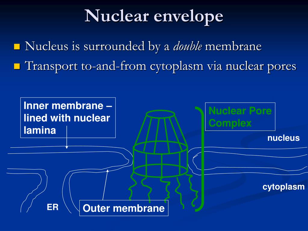

The first hypothesis posits that changes in nuclear shape alter the rigidity of the nucleus this could be beneficial for cells that need to squeeze through tight spaces, but deleterious to cells that are under mechanical duress. In either case, it is still not entirely clear how nuclear shape affects function, although two main hypotheses exist. In some cases, however, the shape of the nucleus is altered by forces that act from the cytoplasm. But what determines nuclear shape, and how does shape affect function? In many cell types, altered nuclear shape is due to changes in the nuclear lamina. Moreover, in certain specialized cell types, altered nuclear shape is important for cell function. This, in itself, is hardly remarkable except for the fact that various diseases, as well as aging, are associated with alterations in nuclear shape ( Fig. The nuclei of most cells are either round or oval. Some organisms, such as Aspergillus nidulans, undergo semi-open mitosis, in which the partial disassembly of the NPCs creates large holes in the NE, but the envelope itself does not completely disassemble (for a review, see De Souza and Osmani, 2007). This strategy works in cell types where the centrosome equivalents, known as spindle-pole bodies, are embedded in the NE, allowing microtubules to associate with chromosomes without the need for NE disassembly. In closed mitosis, the NE does not disassemble and chromosome segregation takes place entirely within the confines of the nucleus. At the end of open mitosis, the NE reassembles around the two segregated DNA masses to form the two daughter nuclei. In open mitosis, the NE disassembles early in mitosis, allowing microtubules that emanate from cytoplasmic centrosomes to contact the chromosomes and promote their segregation (reviewed by Prunuske and Ullman, 2006). Open mitosis occurs in most eukaryotic cells, whereas closed mitosis occurs in certain species of fungi. Two main strategies have evolved to successfully carry out this task: open mitosis and closed mitosis ( Fig. In addition to their location at the nuclear periphery, lamins are also present in the nucleoplasm ( Dechat et al., 2008).ĭuring mitosis, a parent cell gives rise to two daughter cells, each with its own nucleus. This domain is missing in lamin C and is normally removed by proteolytic cleavage in lamin A. Lamin A and the two lamin Bs have a C-terminal domain that is lipid modified (farnesylated), thereby promoting their attachment to the inner nuclear membrane. There are also two major B-type lamins, lamin B1 and lamin B2, each encoded by its own gene. In mammals, there are two major A-type lamins, lamin A and lamin C, which are generated by alternative splicing of the LMNA gene. The nuclear lamina is made predominantly of intermediate filaments called lamins, of which there are two main types: type A and type B (for a review, see Dechat et al., 2008). In metazoans, the NE also contains a meshwork of proteins, which are collectively called the nuclear lamina, that underlies the inner nuclear membrane and interacts with portions of the chromatin. The outer nuclear membrane, which is continuous with the ER, connects with the inner nuclear membrane at the curved membrane regions that surround each NPC.

1) is made of a double membrane that is perforated by nuclear-pore complexes (NPCs). Rather than the insoluble nuclear lamina, the GV content, which is dissolved in the cytoplasm with the onset of oocyte maturation, influences the characteristics and size of transferred nuclei.Most eukaryotic cells have a single nucleus in which a nuclear envelope (NE) separates the chromosomes from the cytoplasm. Our results showed that the removal of the GV nuclear envelope with attached chromatin and chromatin-bound factors does not substantially influence the size of the remodelled nuclei in reconstructed cells and that their nuclear envelopes seem to have normal parameters. In this study, we investigated what other GV components might be necessary for the formation of normal-sized pseudo-pronuclei (PNs). Till date, the only nuclear sub-structure experimentally demonstrated to be essential is the oocyte nucleolus (nucleolus-like body, NLB). However, the nucleus is a complex structure and exactly what nuclear components are required for a successful nucleus remodelling and reprogramming is unknown. It has been shown that the oocyte nucleus (germinal vesicle – GV) components are essential for a successful remodelling of the transferred nucleus by providing the materials for pseudo-nucleus formation. The process of reprogramming/remodelling is, however, only partially characterized. Differentiated nuclei can be reprogrammed/remodelled to totipotency after their transfer to enucleated metaphase II (MII) oocytes.


 0 kommentar(er)
